
There are many different tools physicians use to identify problems in the heart and vascular system. Depending on the symptoms and other health conditions a patient has, your primary care provider, cardiologist or vascular specialists will likely order different tests to help diagnose the problem so they can create the right treatment plan. These tests are performed by highly trained staff and interpreted by board-certified cardiologists and vascular specialists, depending on the test. Results are then part of the patient’s electronic medical record, which can be accessed through MyChart.
Some of these tests are performed in physician offices, some in all of Riverside’s acute care hospitals, and some are only performed at Riverside Regional Medical Center in Newport News, VA. Your provider or the individual scheduling your test will provide directions and any needed instructions for your test. You can always check MyChart for the address and instructions as well.
Different lab tests might be run depending on the symptoms and other health issues. Some tests are ordered as outpatients. For example, labs can determine levels of cholesterol or look for indicators of heart failure or inflammation, which can indicate a problem. Different blood tests may be ordered in the emergency department to check the blood for signs of damaged heart muscle, such as during a heart attack.
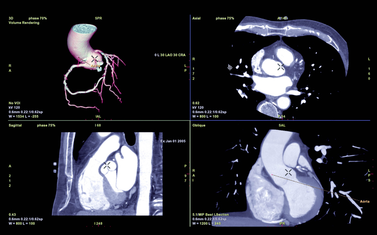
Your provider may order a CT Angiogram scan to get a 3D view of the heart and surrounding blood vessels. Specific dye, called contrast, is injected through an IV and highlights the vessels on the CT image. This will show any blockages or narrowing, and it can help also help evaluate the valves inside the heart.
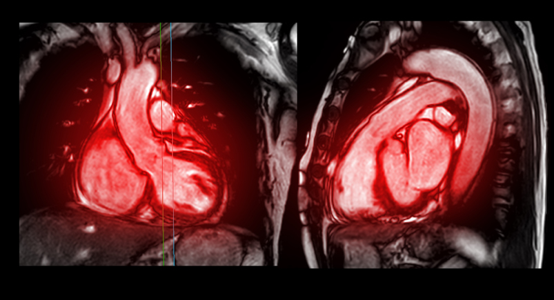
Cardiac Magnetic Resonance Imaging (MRI) is a specific type of technology that takes high quality images of the heart, valves and major vessels utilizing magnetic waves instead of radiation. It can also show plaque build-up and blockages within the blood vessels, as well as identify damaged tissue from cardiovascular disease or a heart attack.
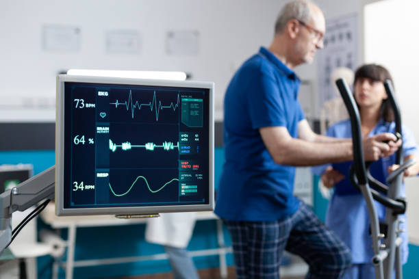
These tests are ordered as a way for your doctor to see how your heart works at rest and when it is working really hard. Usually patients wear the heart monitor leads for an EKG so the heart’s activity can be measured at rest and when “stressed” by the exercise. Blood pressure is also regularly checked during the test. Patients who are unable to do the exercise can be given a medication that causes their heart to act like it does when it is stressed.
Stress tests are often done in conjunction with echocardiography, also called a cardiac echo, to allow the physician to not only see the heart’s electrical activity during the test but to also get a visual image of the heart as it pumps. Like a regular stress tests, cardiac echo tests, this visualization is done with ultrasound technology.
Another common form of stress tests is a nuclear stress test. With this version, the patient is given a small amount of a radioactive tracer substance through an IV. Then they get a PET (positron emission tomography) scan that creates an image of the heart and how the blood moves through it based on where the tracer has gone in the heart. After a period of exercise the patient receives another dose through the IV, and another image is taken so the physician can compare the heart at rest and when stressed. Nuclear stress tests can also go by the more formal names of Cardiac PET, Cardiac SPECT or Myocardial Infusion Pumping Test.
Stress tests [LINK: https://www.riversideonline.com/patients-and-visitors/healthy-you-blog/blog/s/stress-tests ] usually take about an hour for the whole appointment, but the exercise part is usually less than 15 minutes.
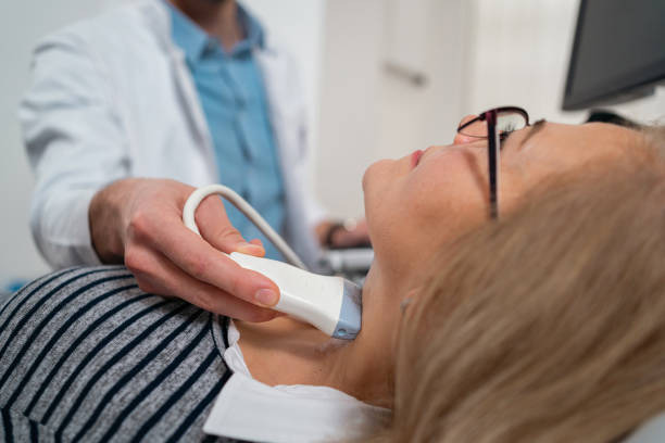
This test uses ultrasound technology to examine the carotid artery, which is how blood gets to our brain. Carotid ultrasounds allow physicians to examine the thickness of the artery’s wall as well as see how blood flows through it. It can identify when the carotid artery is blocked or too narrow, which would increase the risk of stroke. This is noninvasive and involves gel being applied to the neck area and an ultrasound wand pressed to the skin to take the images inside the neck. This test can be performed by highly trained staff, Advanced Practice Providers or physicians, and is read by our board-certified vascular specialists.
This noninvasive test is used to identify, locate and measure the build-up of calcified plaque deposits in the coronary arteries. This results in a calcium score, or coronary artery calcium score, that is used to evaluates a patient’s risk of coronary heart disease.
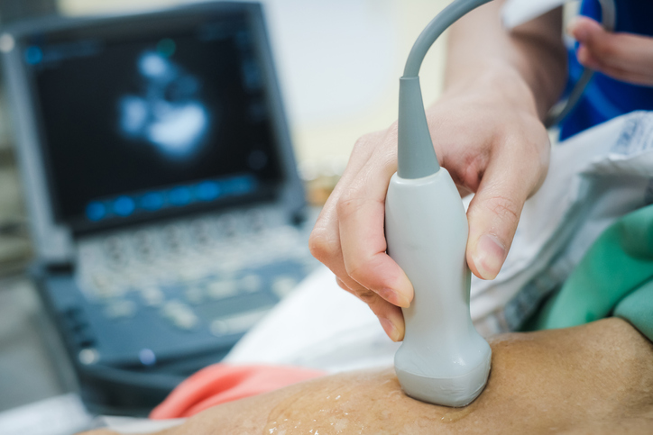
Using the same technology as the ultrasound used to see an unborn baby, the machine uses sound waves to create a live image on the screen. These ultrasound images show the size and shape – as well as the function and efficiency – of the heart muscle. Gel is applied to the skin over the heart, and a device that looks like a wand is pressed to the skin. This painless, noninvasive procedure is performed by highly trained technologists and interpreted by Riverside’s cardiologists.
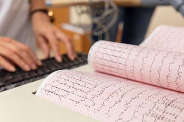
This is a fast and painless procedure that checks for problems with the electrical activity of the heart. An EKG translates the heart’s electrical activity into lines on paper. The spikes and dips on the paper are called waves. From these lines, the cardiologists and other qualified clinicians can take a snapshot of the heart’s rhythm and determine if certain areas (but not all) of the heart muscle have been damaged.
This test is done when someone has an irregular heartbeat to create a detailed map of the heart’s electrical pathway. It is a procedure done in the cardiac catheterization lab by the cardiac electrophysiologists. This can help determine treatments for irregular heartbeats (arrythmias) or establish someone’s risk of cardiac arrest. It is often done immediately prior to an ablation procedure to establish the precise location of the tissue to be ablated.
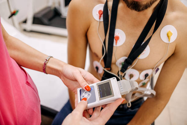
These are all versions of monitors that record someone’s heart rhythm for a long period of time, varying from 24 hours to a month. Patients wear the monitor under their clothes and the monitors record their heart rhythms while they go about their normal life. With some versions patients press a button or record when they notice symptoms so the information can eventually be matched to the rhythm at that time. These monitors are not invasive.
A MUGA scan is a nuclear imaging test to create a video of the pumping heart, highlighting how the blood moves through the lower chambers (or ventricles). This test allows your physician to determine a patient’s ejection fraction (EF). EF measures the percent of blood in the ventricles (lower chambers) that the heart pumps with each heartbeat. The MUGA scan can be done at rest and also in conjunction with a stress test.
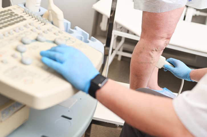
Ultrasound technology can be used on the vascular system throughout the body, including the veins and arteries in arms and legs. This allows physicians to see the health and function of the vascular system. This can include seeing if there are any narrowed areas or blockages. Depending on the location of these issues, it could put someone at risk for blood flow issues in the extremities, such as hands and feet. Or, in other locations, these problems can cause heart attacks and strokes. This is a noninvasive test and involves having gel applied to the skin, and then the ultrasound wand is pressed over the gel to capture the images needed.